A critically important feature: the brain lives in a thick bony mass, the skull, and consequently lacks direct access to the external world.
The brain is a remarkable organ. It receives information from the body through sensing organs like our eyes, ears, skin, and lungs. I would also add the microbiome as the last human sense organ to be studied.
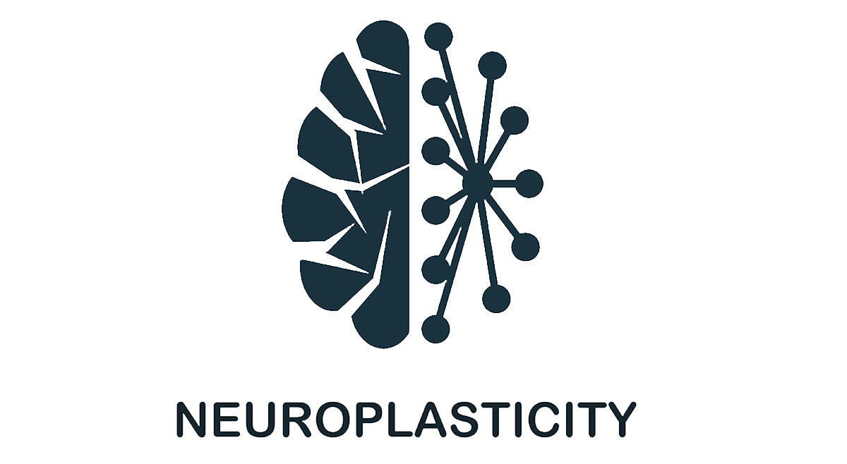
I would like to briefly summarise a few chapters from a booklet I am writing on neuroplasticity, learning, and resilience. My proposal is that inflammation is involved in both normal and aberrant synaptic pruning.
Synaptic function plays a fundamental role in normal brain development. A synapse is a specialised junction between two neurons or between a neuron and an effector cell. It is the connection point for electrical signals (action potentials) to pass from one neuron to the next, allowing neural communication throughout the nervous system. Synapses are highly regulated by various physiological processes, modulating their activity further by neurotransmitters such as dopamine, noradrenaline, and serotonin. By understanding how synapses work and operate, as educators and parents, we can influence and nurture the maturing brains of our children.
In response to this varied sensory input, the infant’s brain engages in massive synaptic growth during the first two years of life. The synaptic connections in an infant’s brain increase exponentially; up to two million synapses occur each second!
By age two, they have over one hundred trillion of these synaptic connections, double that of a fully-grown adult. After this, synaptic pruning occurs, where synapses decrease dramatically. Those actively used synapses are thought to be maintained, and those seldom used are lost. The outcome of this pruning results in a brain where neural circuits, for better or worse, are embedded in the subconscious.
The mechanism of synaptic pruning remains poorly understood, although the innate immune system appears to be involved. An idea that I proposed some 25 years ago was that the innate immune system (in the form of dendritic cells) would sense the pathogen environment and respond by producing pro-inflammatory cytokines (DC1) or anti-inflammatory cytokines (DC2). I had no idea then that the nervous system also regulated this process.
Of particular interest is that the resident phagocytic cells in the brain, the microglia, are similarly activated to release pro-inflammatory cytokines (M1) or anti-inflammatory cytokines (M2). These cells are thought to maintain organ homeostasis by clearing out cellular debris, such as those that die through apoptosis.
The burning questions are what drives the maturation of microglial cells down either of these two pathways and what features of a synapse give the go-ahead for the microglial cell to phagocytose and remove the glial cells.
It is well-established that infection can drive this polarisation into M1 or M2 phenotypes. However, in recent years it is becoming clear that there is neural control, also. For example, the parasympathetic nervous system (flight or fight response) can drive the pro-inflammatory response, including heart rate increase and hypertension. In contrast, the sympathetic nervous system promotes calmness and an anti-inflammatory milieu of cytokines.
But why are active neuronal circuits preserved from synaptic pruning? I propose a model where action potentials increase the synaptic expression of these tag(s) and protect the synapse from deletion by microglial cells. Active synapses will express this molecule highly, and inactive synapses will not. A corollary to this idea is that the downregulation of this tag on active synapses could easily lead to aberrant pruning.
However, in addition to action potentials, other extracellular factors, such as pro-inflammatory cytokines, regulate expression.
One such molecular tag is a glycoprotein CD47. This molecule is expressed on several cell types and protects from phagocytosis. Interestingly, CD47 is expressed on synapses, and action potentials likely regulate its expression. In addition, proinflammatory cytokines down-regulate CD47 expression and make the synapse susceptible to deletion. These normal processes underpin what we think of as creating normal brain architecture.
This new model informs parents, educators, and the government on how to create a brain that thrives in the cultural environment we have decided on.
Toxic stress promotes a predictable parasympathetic nervous system activation, leading to the chronic production of proinflammatory cytokines such as IL-6 and TNF. This then leads to downregulating of the CD47 protective tag, and as a consequence, we see abnormal synaptic pruning, which can lead to culturally neuroatypical outcomes.
On the other hand, promoting well-being through Gratitude, Exercise, Nutrition, and meaningful relationships promotes the activation of the sympathetic nervous system and the generation of M2 Microglial cells.
This model suggests that soon, we should have a metric that measures resilience. Worryingly, it also suggests that school readiness is a cultural illusion the government chooses. In a later chapter, I will explain why this evidence suggests that resilience is still a few years away for a population of children and that formal education should start at age 7.